|
Diagnostic Imaging
Equipment |
|
|
|
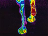
Thermography |
|
Thermal imaging uses
new advanced infrared technology to detect heat and inflammation. It
is an excellent tool for those tough lameness's and soft tissue
injuries. We have been using thermal imaging since 1988. At times
we may cool the horse or an area with a cold water bath / hose then watch
for heat to radiate out from deeper structures. Click on the link
below for more information and case studies.
|
|
|

|
|
Click
here for more information on thermography
|
|
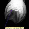
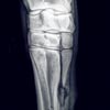
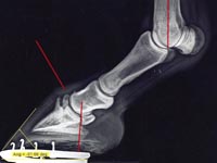
Radiography
|
|
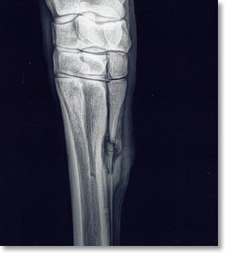
Advanced digital radiology equipment can
be used to diagnose a mulitude of bone-related abnormalities that result in
lameness problems. Common issues range from hock arthritis, coffin bone
remodeling, kissing spine lesions to navicular disease. With our state of
the art equipment we can determine ideal coffin bone alignment to work with
your farrier for corrective shoeing. Click on the link below for more
information and case studies. |
|
|

|
|
Click here
for more
information on radiography
|
|
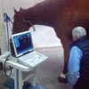
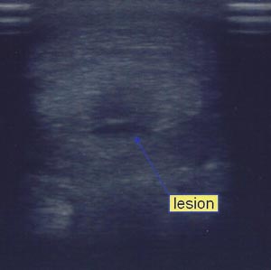
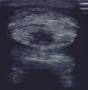
Ultrasonography
|
|
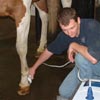
State of the art ultrasonograpy equipment
may be used to diagnose lesions in many structures throughout the horse.
From tendon and ligament lesions to displaced intestines and lung pathology.
|
|

|
|
Endoscopy
|
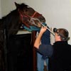
Endoscopy
enables visualization of several structures in the horse. We are
equipped with several different sized scopes to complete a variety of
procedures. Some examples of this include evaluation of the upper
respiratory tract for coughs and airway noise during exercise. We
can also visualize the stomach for gastric ulcer diagnosis.
|
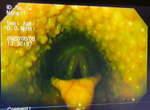
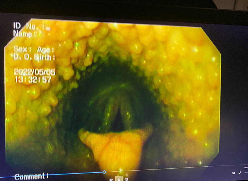
Hyperactive, proliferative tonsilar
tissue in a 2 year old TB |
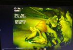
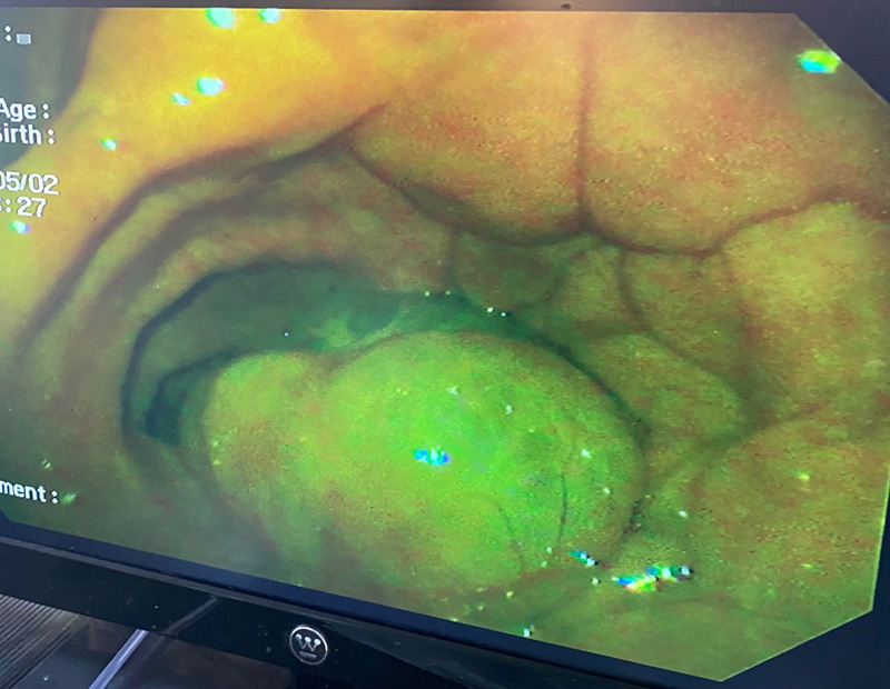
Severe fungal infection (mycosis)
of the guttural pouch
|

Examination of the stomach
pylorus via gastroscopy |

Large uterine cyst via hysteroscopy
|
|
Uterine cyst ablation.mov |